Smash a melon, NOT my Boob!
Do Mammograms CAUSE Cancer?
“Routine Mammograms do not save lives, the research is clear.”
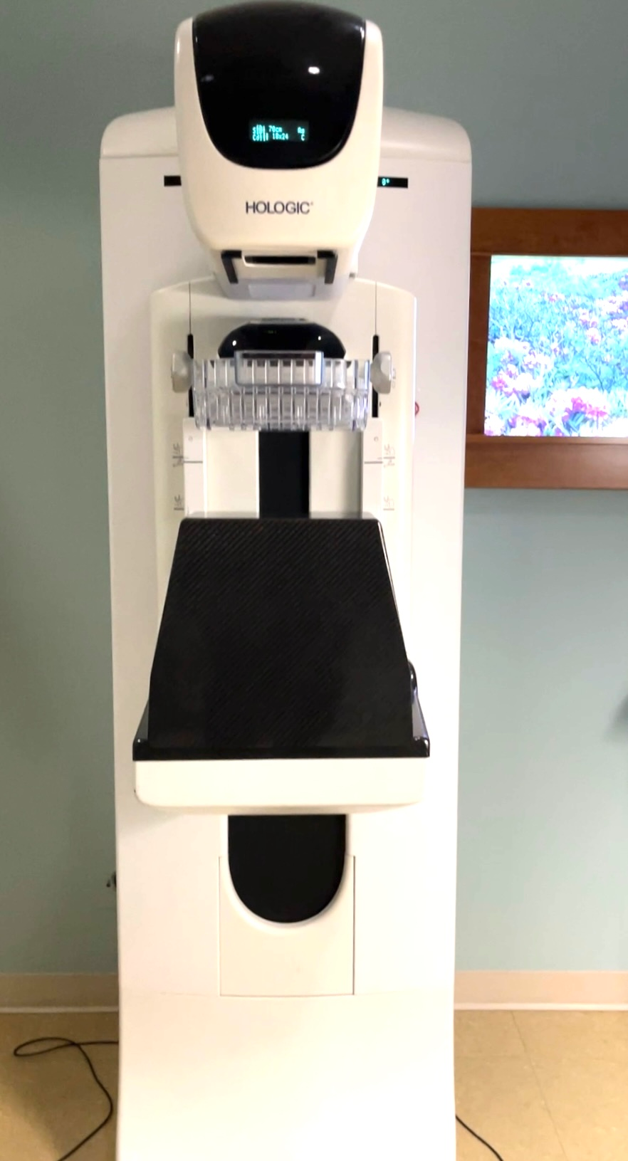
Mammography is a type of X-ray imaging that utilizes ionizing radiation and breast compression. Unlike bone, which X-rays were designed to image, breast tissue is soft and absorbs radiation poorly. To enhance contrast between fat and glandular tissue, mammograms use lower-energy X-rays (20–30 keV). However, this increases radiation absorption in the breast, a tissue especially vulnerable to radiation damage. Since exposure is cumulative, repeated mammograms carry a risk of contributing to cancer development.
The U.S. National Cancer Institute states that while mammograms use small doses, “repeated x-rays have the potential to cause cancer,” so patients should discuss the need for each exam. Cancer.gov

Radiation from Mammograms
Radiation Induced Cancer & Death
Annual mammography screening carries a measurable risk of radiation-induced breast cancer and related deaths. The earlier women begin regular mammograms, the greater their cumulative risk over time. Women with higher radiation exposure or those requiring additional imaging, such as women with larger or dense breasts, face an increased risk compared to others.
“There is no safe level of exposure and there is no dose of radiation so low that the risk of a malignancy is zero” -Dr. Karl Z. Morgan, dubbed the father of Health Physics
Radiation-Induced Breast Cancer Incidence and Mortality from Digital Mammography Screening: A Modeling Study explains, on average, annual screening of 100,000 women aged 40 to 74 years was projected to induce 125 breast cancers (95% confidence interval [CI]=88–178) leading to 16 deaths (95% CI=11–23), relative to 968 breast cancer deaths averted by early detection from screening. Women exposed at the 95th percentile were projected to develop 246 radiation-induced breast cancers leading to 32 deaths per 100,000 women. Women with large breasts requiring extra views for complete breast examination (8% of population) were projected to have higher radiation-induced breast cancer incidence and mortality (266 cancers, 35 deaths per 100,000 women), compared to women with small or average breasts (113 cancers, 15 deaths per 100,000 women).
Both standard 2D mammograms and 3D tomosynthesis (DBT) expose the breast to ionizing radiation, which can increase the risk of cancer over time. DBT takes multiple images from different angles, and while each individual image uses less radiation than a single 2D mammogram, the total radiation exposure is higher, especially when DBT is combined with traditional 2D images as is the case typically. Using synthetic 2D images can reduce the dose somewhat, but exposure still occurs and varies depending on breast size, density, and imaging technique.
What’s the risk? John W. Gofman’s 1998 analysis, “Mammography: An Individual’s Estimated Risk that the Examination Itself Will Cause Radiation-Induced Breast Cancer,” provides estimates of cancer risk associated with mammography based on radiation exposure. More recent research about “Mammography-induced cancers” by Daniel Corcos is discussed in video here.
3d Mammograms
digital breast tomosynthesis (DBT)
Since FDA approval in 2011, 3D mammography, or digital breast tomosynthesis (DBT), has been heavily marketed as a superior alternative to standard 2D mammography by manufacturers, must-have upgrade, branded as “genius 3D” or “smart 3D” technology“. Hospitals and clinics advertise it as “safer,” “clearer,” or “more accurate” without always disclosing that it involves increased radiation dose compared to 2D and the financial incentives which drive agressive marketing.
The combination of Medicare coverage, state mandates, and higher reimbursement rates has created a strong financial incentive to promote 3D mammography (DBT). In 2014, Medicare began reimbursing DBT when performed with standard 2D mammography, and by 2021, 17 states required private insurers to cover DBT without cost-sharing. Radiology providers can earn more for performing DBT than standard 2D mammograms, which has driven aggressive marketing of DBT as a “must-have” upgrade. These financial pressures can influence clinics to prioritize adoption and patient uptake, overshadowing honest, evidence-based discussions about whether DBT is medically necessary for each individual, leading to screening decisions that are motivated by profit.
Early Screening
Early Screening means more Radiation
In 2024, U.S. breast cancer screening policy shifted significantly. The USPSTF lowered the recommended age for biennial mammograms from 50 to 40 for women at average risk. For BRCA1 and BRCA2 mutation carriers, guidelines recommend earlier, more frequent monitoring, with mammograms starting at age 30 (American Cancer Society). Yet this also increases cumulative lifetime radiation and therefore cancer risk. In addition, U.S. facilities also began notifying women of their breast density with encouragement to pursue additional—higher radiation—imaging such as 3D mammography. Learn more about Dense Breasts and Underdiagnosis here.
“The risk of breast cancer almost doubled after 15 years of screening. Additional cancers began to occur less than 6 years after mammography. These results are evidence that X-ray-induced carcinogenesis, rather than overdiagnosis, is the cause of the increase in breast cancer incidence.” – Daniel Corcos
Our Stories
My Mom was diagnosed about same time and did the same procedure. They gave her so much radiation that she developed a benign tumor in her esophagus that was directly caused by the radiation. It ruptured her esophagus and she had to get her esophagus removed. The side effects and consequences of Radiation are real and some are catastrophic. This was done at a major medical hospital. They should not be using radiation for DCIS unless it is high grade and only if a woman is informed and educated about possible life long side effects. It should be taken more seriously. But, women are not usually informed prior to having it done.
Smash a melon, mot my boob!
Disclaimer
The information provided on Smash a Melon, NOT My Boob! is for educational and awareness purposes only and is not a substitute for professional medical advice, diagnosis, or treatment. We are not medical professionals.
Always seek the advice of a qualified healthcare provider regarding any medical condition, screening decision, or treatment option.
Connect with us today and join our challenge!
Subscribe
Join the conversation
Help us smash the silence.